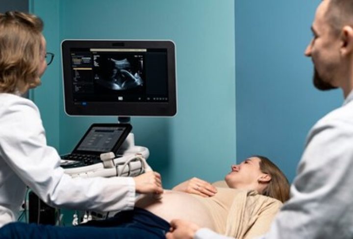
It is also known as fetal neurosonogramis, designed to assess the developing brain and central nervous system of a fetus during pregnancy. It plays a crucial role in prenatal care by providing valuable insights into the health and development of the unborn baby. Typically performed between 18 and 22 weeks of gestation and is a safe and non-invasive procedure that utilizes sound waves to create detailed images of the fetal brain, spinal cord, and surrounding structures. Early detection can significantly improve outcomes for both the baby and the mother, as it enables Doctors to monitor and timely intervention.
Preparing for Fetal Neurosonography
- Fasting: Follow instructions provided by your doctor such as fasting or drinking water before the procedure.
- Comfortable Clothing: Wear loose-fitting and comfortable clothing to your appointment, as you may need abdominal ultrasound.
Procedure for Fetal Neurosonography
- Preparation: The expectant mother may be asked to drink water before the procedure to fill the bladder, providing better visualization of the fetus.
- Positioning: The mother will lie on an examination table, and a gel will be applied to her abdomen to help transmit sound waves.
- Ultrasound Probe: It also known as a transducer, to transmit high-frequency sound waves into the abdomen.
- Image Capture: The sound waves bounce off the fetal structures and create detailed images of the developing brain, spinal cord, and surrounding structures on a monitor.
- Analysis: The Doctor will analyze the images in real-time, looking for any abnormalities or developmental issues and discuss the findings with parents.
Benefits of Fetal Neurosonography
- Early Detection: It enables the early detection of abnormalities in the fetal brain and central nervous system, allowing for timely interventions and management.
- Reassurance: Seeing detailed images of the developing fetal brain and central nervous system can provide reassurance to expectant parents about the health and well-being of their baby.
- Improved Outcomes: Early detection of abnormalities can lead to improved outcomes for both the baby and the mother by facilitating closer monitoring of the pregnancy and intervention if necessary.
- Monitoring High-Risk Pregnancies: It is particularly valuable in monitoring high-risk pregnancies, where there may be a greater likelihood of fetal abnormalities or complications.
Fetal neurosonography is a vital tool in prenatal care, offering early detection of abnormalities, informed decision-making, and reassurance to expectant parents. Its ability to improve outcomes through timely interventions and support multidisciplinary collaboration makes it indispensable in modern obstetrics.


